ACL Injuries
When it comes to knee injuries, one of the most devastating is an ACL sprain. Whether you’re a professional athlete or a weekend warrior, this injury can leave you sidelined for months to over a year. Lets break down an ACL sprain below.
ACL Sprain Guide
Anatomy
How does it happen
How do you diagnosis it
Treatment
ACL Anatomy
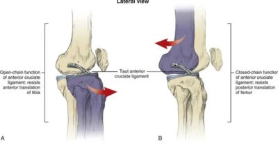
The ACL is the most important ligament in the knee for stability (source). It prevents anterior translation of the tibia. This is a function the ACL shares with the hamstring group. This will be relevant further down in this post.
The tibial nerve and middle genicular artery innervate the ACL respectively. This blood vessel plays a major role in recovery as well as the visible symptoms as the swelling post injury can be made worse if the artery is damaged.
In any knee injury, if there is a loss of proper foot control, primarily the ability to dorsiflex, go to the ER immediately. This is indicative of nerve damage and needs emergent attention.
As with all ligaments, there are 3 grades of sprains:
Grade 1 – Stretching of the tissue with minimal to no tearing
Grade 2 – Moderate tearing of the fibers
Grade 3 – Full rupture of the ligament
How Does An ACL Sprain Happen?
The most common way to tear an ACL is is a non contact injury where the knee buckles. This usually applies a valgus force at the knee which can tear the ACL, along with the MCL and meniscus in several cases.
The other way is forceful valgus force. This usually results from contact sports and another athlete falls/gets thrown into the outside of the knee. Valgus force goes to the knee, tearing the ACL.
Prevention for an ACL tear lies in a knee brace for contact sports. The brace will limit the force applied to the knee, decreasing injury severity. In terms of preventing a non contact injury, there needs to be a focus on strength and plyometric training to increase force tolerance at the knee.
Additionally, playing surface is a large factor regarding outdoor competition. Injury rates on turf are substantially higher than on grass. If an athlete’s foot is stuck in the ground when force is applied, usually the grass will give out and the athlete slips. Turf does not give out, therefore the force travels up and the soft tissue gives out.
How Is An ACL Sprain Diagnosed?
In terms of diagnosis, a Lachman’s test is the gold standard for a clinical exam. A good clinician can tell if the ACL is torn before an athlete gets off the field. In this case the MRI confirms what the exam found as opposed to being the primary assessment. This test looks for anterior translation. If this motion exists, the ACL is damaged/torn.
Another ACL test is the anterior drawer. This looks for the same movement, however, the hamstrings get in the way of this test’s accuracy from guarding or spasm. As the hamstrings resist anterior translation as well, this test is not as reliable.
With all evaluations, the ultimate gold standard for soft tissue damage is an MRI. With the knee, and ACL specifically, the clinical exams are good and reliable in the hands of a good clinician. As such, the MRI confirms the clinical exam.
How Do You Treat An ACL Sprain?
The type of treatment needed to rehab an ACL has some variation, but generally it follows the same guidelines:
Grade 1 and 2 sprain – Conservative rehab
Grade 3 in a non athletic population – Conservative rehab (lotta nuance here)
Grade 3 in an athletic population – Surgery
Conservative Rehab
It is very possible and quite normal to live life without an ACL. The goals of rehab are to restore ROM, build up strength, return to normal activities/plyometrics. The only variable to this is how long each grade will take to heal/return to function. A grade 3 tear will never heal. Obviously there are many variables that can determine the healing times (age, fitness level, strength etc).
Grade 1 – Several weeks
Grade 2 – 2-3 months
Grade 3 – 6 months plus
Surgery
There are 3 commonly used surgical techniques for an ACL surgery
Hamstring Tendon Graft
A portion of the hamstring tendon is harvested from the back of the knee and then grafted into the spot where the ACL once was. This process is call ligamentization. The body is capable of turning a tendon into a ligament.
The main concern with this graft is that the hamstrings share a function with the ACL if you recall from earlier. Both structures resist anterior translation. By harvesting the hamstring, the tissue is made weaker as part of it is removed.
Patella Tendon Graft
This is the gold standard for ACL tears. This graft is a BTB (bone-tendon-bone). What happens is the middle third of the patella tendon is harvested. Part of the bone at the femur and the tibia comes with the tendon.
When this is grafted to replace the ACL, bone is fused to bone. This is allows for a faster healing time as bone heals faster than soft tissue. This leaves the hamstrings intact as well which generally leads to better outcomes.
Cadaver Graft
This process takes an ACL from a cadaver and then grafts it into the knee. This type of graft has a high chance of failure. One reason being that the body could simply reject the foreign tissue. I have never had an athlete undergo this type of graft. It is far from the ideal option. The risk of failure is far too great when there are much better options.
In summary, an ACL tear is a long term injury with many factors that can determine outcomes. As devastating as the injury is, we have gotten quite good at fixing them and restoring knee function.
If you have an ACL injury, or any knee injury you need help with, reach out to us here


